РЕЗОЛЮЦІЯ: Громадського обговорення навчальної програми статевого виховання
ЧОМУ ФОНД ОЛЕНИ ПІНЧУК І МОЗ УКРАЇНИ ПРОПАГУЮТЬ "СЕКСУАЛЬНІ УРОКИ"
ЕКЗИСТЕНЦІЙНО-ПСИХОЛОГІЧНІ ОСНОВИ ПОРУШЕННЯ СТАТЕВОЇ ІДЕНТИЧНОСТІ ПІДЛІТКІВ
Батьківський, громадянський рух в Україні закликає МОН зупинити тотальну сексуалізацію дітей і підлітків
Відкрите звернення Міністру освіти й науки України - Гриневич Лілії Михайлівні
Представництво українського жіноцтва в ООН: низький рівень культури спілкування в соціальних мережах
Гендерна антидискримінаційна експертиза може зробити нас моральними рабами
ЛІВИЙ МАРКСИЗМ У НОВИХ ПІДРУЧНИКАХ ДЛЯ ШКОЛЯРІВ
ВІДКРИТА ЗАЯВА на підтримку позиції Ганни Турчинової та права кожної людини на свободу думки, світогляду та вираження поглядів
- Гідрологія і Гідрометрія
- Господарське право
- Економіка будівництва
- Економіка природокористування
- Економічна теорія
- Земельне право
- Історія України
- Кримінально виконавче право
- Медична радіологія
- Методи аналізу
- Міжнародне приватне право
- Міжнародний маркетинг
- Основи екології
- Предмет Політологія
- Соціальне страхування
- Технічні засоби організації дорожнього руху
- Товарознавство продовольчих товарів
Тлумачний словник
Авто
Автоматизація
Архітектура
Астрономія
Аудит
Біологія
Будівництво
Бухгалтерія
Винахідництво
Виробництво
Військова справа
Генетика
Географія
Геологія
Господарство
Держава
Дім
Екологія
Економетрика
Економіка
Електроніка
Журналістика та ЗМІ
Зв'язок
Іноземні мови
Інформатика
Історія
Комп'ютери
Креслення
Кулінарія
Культура
Лексикологія
Література
Логіка
Маркетинг
Математика
Машинобудування
Медицина
Менеджмент
Метали і Зварювання
Механіка
Мистецтво
Музика
Населення
Освіта
Охорона безпеки життя
Охорона Праці
Педагогіка
Політика
Право
Програмування
Промисловість
Психологія
Радіо
Регилия
Соціологія
Спорт
Стандартизація
Технології
Торгівля
Туризм
Фізика
Фізіологія
Філософія
Фінанси
Хімія
Юриспунденкция
MUCOSAL-ASSOCIATED LYMPHOID TISSUE
The mucous membranes lining the digestive, respiratory, and urogenital systems have a combined surface area of about 400 m2 (nearly the size of a basketball court) and are the major sites of entry for most pathogens. These vulnerable membrane surfaces are defended by a group of organized lymphoid tissues mentioned earlier and known collectively as mucosal-associated lymphoid tissue (MALT).Structurally, these tissues range from loose, barely organized clusters of lymphoid cells in the lamina propria of intestinal villi to well-organized structures such as the familiar tonsils and appendix, as well as Peyer’s patches, which are found within the submucosal layer of the intestinal lining. The functional importance of MALT in the body’s defense is attested to by its large population of antibody-producing plasma cells, whose number far exceeds that of plasma cells in the spleen, lymph nodes, and bone marrow combined.
The tonsilsare found in three locations: lingual at the base of the tongue; palatine at the sides of the back of the mouth; and pharyngeal (adenoids) in the roof of the nasopharynx (Figure 7). All three tonsil groups are nodular structures consisting of a meshwork of reticular cells and fibers interspersed with lymphocytes, macrophages, granulocytes, and mast cells. The B cells are organized into follicles and germinal centers; the latter are surrounded by regions showing T-cell activity. The tonsils defend against antigens entering through the nasal and oral epithelial routes.
The best studied of the mucous membranes is the one that lines the gastrointestinal tract. This tissue, like that of the respiratory and urogenital tracts, has the capacity to endocytose antigen from the lumen. Immune reactions are initiated against pathogens and antibody can be generated and exported to the lumen to combat the invading organisms. As shown in Figures 8 and 9, lymphoid cells are found in various regions within this tissue. The outer mucosal epithe-lial layer contains so-called intraepithelial lymphocytes (IELs).Many of these lymphocytes are T cells that express unusual receptors (γδT-cell receptors, or γδ TCRs), which exhibit limited diversity for antigen. Although this population of T cells is well situated to encounter antigens that enter through the intestinal mucous epithelium, their actual function remains largely unknown The lamina propria, which lies under the epithelial layer, contains large numbers of B cells, plasma cells, activated TH cells, and macrophages in loose clusters. Histologic sections have revealed more than 15,000 lymphoid follicles within the intestinal lamina propria of a healthy child. The submucosal layer beneath the lamina propria contains Peyer’s patches, nodules of 30–40 lymphoid follicles. Like lymphoid follicles in other sites, those that compose Peyer’s patches can develop into secondary follicles with germinal centers.

Figure 7. Three types of tonsils. (a) The position and internal features of the palatine and lingual tonsils; (b) a view of the position of the nasopharyngeal tonsils (adenoids). [Part b adapted from J. Klein, 1982, Immunology, The Science of Self-Nonself Discrimination, © 1982 by John Wiley and Sons, Inc.]
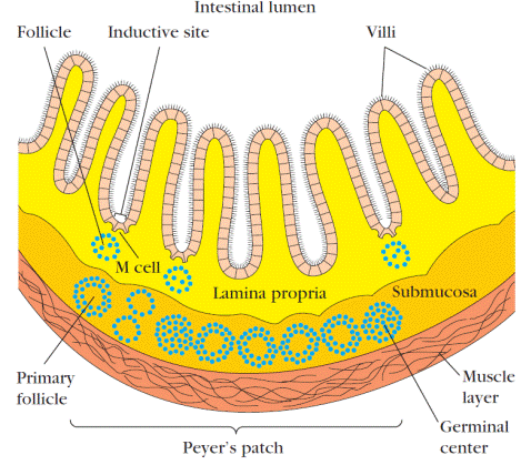
Figure 8. Cross-sectional diagram of the mucous membrane lining the intestine showing a nodule of lymphoid follicles that constitutes a Peyer’s patch in the submucosa. The intestinal lamina propria contains loose clusters of lymphoid cells and diffuse follicles.
The epithelial cells of mucous membranes play an important role in promoting the immune response by delivering small samples of foreign antigen from the lumina of the respiratory, digestive, and urogenital tracts to the underlying mucosal-associated lymphoid tissue. This antigen transport is carried out by specialized M cells.The structure of the M cell is striking: these are flattened epithelial cells lacking the microvilli that characterize the rest of the mucous epithelium.
In addition, M cells have a deep invagination, or pocket, in the basolateral plasma membrane; this pocket is filled with a cluster of B cells, T cells, and macrophages (Figure 9a). Luminal antigens are endocytosed into vesicles that are transported from the luminal membrane to the underlying pocket membrane. The vesicles then fuse with the pocket membrane, delivering the potentially response-activating antigens to the clusters of lymphocytes contained within the pocket.
M cells are located in so-called inductive sites – small regions of a mucous membrane that lie over organized lymphoid follicles (Figure 9b). Antigens transported across the mucous membrane by M cells can activate B cells within these lymphoid follicles. The activated B cells differentiate into plasma cells, which leave the follicles and secrete the IgA class of antibodies. These antibodies then are transported across the epithelial cells and released as secretory IgAinto the lumen, where they can interact with antigens.
Mucous membranes are an effective barrier to the entrance of most pathogens, which thereby contributes to nonspecific immunity. One reason for this is that the mucosal epithelial cells are cemented to one another by tight junctions that make it difficult for pathogens to penetrate. Interestingly, some enteric pathogens, including both bacteria and viruses, have exploited the M cell as an entry route through the mucous-membrane barrier. In some cases, the pathogen is internalized by the M cell and transported into the pocket. In other cases, the pathogen binds to the M cell and disrupts the cell, thus allowing entry of the pathogen. Among the pathogens that use M cells in these ways are several invasive Salmonella species, Vibrio cholerae, and the polio virus.

Figure 9. Structure of M cells and production of IgA at inductive sites. (a) M cells, located in mucous membranes, endocytose antigen from the lumen of the digestive, respiratory, and urogenital tracts. The antigen is transported across the cell and released into the large basolateral pocket. (b) Antigen transported across the epithelial layer by M cells at an inductive site activates B cells in the underlying lymphoid follicles. The activated B cells differentiate into IgA-producing plasma cells, which migrate along the submucosa. The outer mucosal epithelial layer contains intraepithelial lymphocytes, of which many are CD8+ T cells that express γδ TCRs with limited receptor diversity for antigen.
Читайте також:
- Cutaneous-Associated Lymphoid Tissue
- Primary Lymphoid Organs
- Secondary Lymphoid Organs
- Staining of sections of muscular tissue with iron hematoxylin demonstrated cross striation. What other morphological features is cardiac muscle characterized by?
- What tissue takes an active part in heat production in the newborn organism?
- What type of connective tissue contains a large number or melanocytes?
- What type of tissue is characterized by the polar differentiation of the cells?
| <== попередня сторінка | | | наступна сторінка ==> |
| LYMPH NODES | | | Cutaneous-Associated Lymphoid Tissue |
|
Не знайшли потрібну інформацію? Скористайтесь пошуком google: |
© studopedia.com.ua При використанні або копіюванні матеріалів пряме посилання на сайт обов'язкове. |